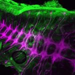
First Prize: Dr. Anna Franz, School of Biochemistry, Faculty of Medical Sciences, University of Bristol.
The beauty and complexity of the central nervous system of a grasshopper embryo is revealed by anti-HRP staining (coloured purple). Other tissues are stained with anti-acetylated tubulin (coloured green).
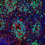
2nd prize: Dr. Patrick Ovando-Roche, Institute of Reproductive and Developmental Biology, Faculty of Medicine, Imperial College London
Neural differentiation of human embryonic stem cells (hESCs): Following neural induction of hESCs, cells form neural rosettes structures to give rise to neural progenitors. Neural rosettes can be spotted by their rosette-like conformation and positive co-expression of pax6 (green) and nestin (red). Cell nuclei were counterstained with DAPI (blue).
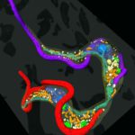
3rd prize: Dr. Louise Hughes, Bio-Imaging Unit, Department of Biological and Medical Sciences, Oxford Brookes University, Oxford.
Kissing trypanosomes: The image shows the final stage of cell division for the single-celled parasite, Trypanosoma brucei, with the two daughter cells connected at their posterior end. The image is generated from reconstructed data from serial block face scanning electron microscopy, showing the cells, their flagella (red and purple) and internal organelles.
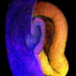
Highly commended: Timothy Grocott, School of Biological Sciences, University of East Anglia, Norwich.
Multi-photon imaging of the embryonic eye: A false-coloured image showing a cutaway through the invaginating lens placode (blue tissue layer) situated within the optic cup (yellow tissue layer) of a 2.5-day chick embryo. The surrounding mesenchyme was removed for clarity, while cell nuclei are labelled red.
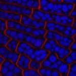
Highly Commended: Dr. Mistianne Feeney, School of Life Sciences, University of Warwick, Coventry
Arabidopsis thaliana embryo cells imaged by confocal microscopy. Long Caption: The plasma membrane of the Arabidopsis thaliana embryo cells is stained with FM4-64 (shown in red) and protein storage vacuoles autofluoresce (shown in blue).