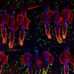
1st Prize: Kif Liakath-Ali
Exquisitely dotted melanocytes of mouse skin revealed by anti-TRP1 staining (Green) and individual hair follicles stained by anti-keratin14 (Red). Blue indicates DAPI staining of cell nuclei and autofluorescent hair shafts. Wholemount immunostaining was carried out on mouse tail epidermis.
Exquisitely dotted melanocytes of mouse skin revealed by anti-TRP1 staining (Green) and individual hair follicles stained by anti-keratin14 (Red). Blue indicates DAPI staining of cell nuclei and autofluorescent hair shafts. Wholemount immunostaining was carried out on mouse tail epidermis.
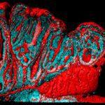
2nd Prize: Alistair Langlands
Small intestinal polyp of an ApcMin mouse. An IMARIS-rendered surface of Phalloidin (red) and nuclei (cyan) show changes associated with early cancer development. Normal structure is lost as the lumen becomes highly folded and epithelial cells start piling on top of one another due to excess proliferation.
Small intestinal polyp of an ApcMin mouse. An IMARIS-rendered surface of Phalloidin (red) and nuclei (cyan) show changes associated with early cancer development. Normal structure is lost as the lumen becomes highly folded and epithelial cells start piling on top of one another due to excess proliferation.
Small intestinal polyp of an ApcMin mouse. An IMARIS-rendered surface of Phalloidin (red) and nuclei (cyan) show changes associated with early cancer development. Normal structure is lost as the lumen becomes highly folded and epithelial cells start piling on top of one another due to excess proliferation.
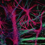
3rd Prize: Joanna Wardyn
This is a confocal image showing intricate structure of astroglial cells (pink, stained with anti GFAP antibody) interconnected with neurons (stained light blue with anti-β III tubulin). Astrocytes are increasingly appreciated as key modulators of neuronal health and function.