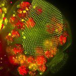
1st Prize: Anna Franz
The head of a Drosophila pupa: The developing compound eye (green) is composed of several hundred simple units called ommatidia arranged in an extremely regular array. The giant polyploidy cells of the fat body (red), the fly equivalent of the mammalian liver and adipose tissue, occupy a big area of the head.
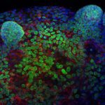
2nd Prize: Ronan Mellin
Confocal image showing a murine colonic epithelial organoid grown in Matrigel (3D culture). Crypt-like projections containing epithelial progenitors can be seen protruding from the spheroid. Stained for DNA with DAPI (Blue), the nuclear envelope with LaminB1 (Green) and the centrosome marker γ-tubulin (Red).
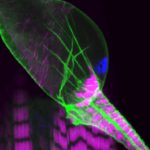
3rd Prize: Helen Weavers
This confocal image shows the intricate structure of the developing fly kidney, which is anchored within the body by attachment to nearby heart muscle. The striking striations of the heart (and body wall muscle beneath) are revealed by labeling Actin (majenta). Cell membranes (green) and nuclei (blue) are also stained.