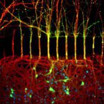
1st Prize: Cristiano Lucci
The image shows primary cortical neurons cultured in these compartmentalised microfluidic chambers. The labelling is for acetylated tubulin in red (identifying all axons), and green for the cell permeable dye calcein, which is only applied on the axonal side of the chambers (top half) and allows the identification of those neuronal cell bodies (bottom half) that have extended axons to the other side of the microfluidic device. Blue staining indicates nuclei labelled with DAPI. The image was taken using a fluorescent microscope at the SLIM facility in the School of Life Sciences.
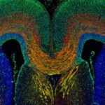
2nd Prize; Anneliese Norris
A section through the embryonic mouse telencephalon at E18.5 showing the L1 expressing corpus callosum (labelled orange) and cells of cortical origin labelled with GFP (green). Section is counterstain with DAPI (blue).
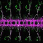
3rd prize tie = Mohammad Moffateh
Beautifully repeated segments of Drosophila melanogaster embryonic nervous system, stage 16/17, ventral view. Stained for Futsch (green, segmented sensory neurons and dispersed cell bodies in the ventral nerve cord) and Ank2-L (magenta, segmented ventral nerve cord and a subset of sensory neurons). Imaged with a Zeiss LSM780 confocal microscope.
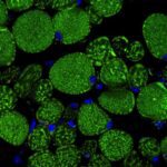
3rd prize tie = Alan Prescott
Confocal image of a transverse section of the rectus muscle of the eye taken from the mito-QC mouse (McWilliams et al., (2016) JCB, 214(3)). Mitochondria express eGFP and mCherry but in lysosomes the eGFP, green fluorescence is quenched. Bright red dots are mitolysosomes. Nuclei are DAPI blue.