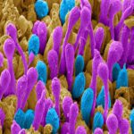
1st Prize: Patrick Ovando Roche
Scanning electron microscopy image of a 16-week-old human retinal organoid generated from pluripotent stem cells using bioreactor technology. Image has been pseudo-coloured to highlight rod (Purple) and cone (Cyan) photoreceptor outer-segments, the cell structures of the retina capable of capturing light and transforming it into vision.
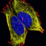
2nd Prize: Lisa Romano
The confocal image shows neuroblastoma cells cultured in a fibronectin coated coverslip, which allowed the formation of focal adhesion structure required during cell migration. The labelling is for vimentin in yellow (cytoskeleton), red for focal adhesion marker vinculin and blue staining for nuclei (DAPI).
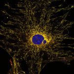
3rd Prize: Alan Prescott Confocal image of a cultured mouse embryo fibroblast from the mito-QC mouse. Mitochondria express both eGFP and mCherry but in lysosomes the eGFP, green fluorescence is quenched. Bright red dots are mitolysosomes. The nucleus is DAPI stained, blue.
Read more about the people behind these images here.