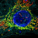
1st Prize: Hope Isobel Needs
Structured Illumination Microscopy (SIM) image of a HeLa cell expressing mScarlet localised to the mitochondrial matrix (red). Mitochondrial membranes are shown in green (MitoTracker Green), cell nuclei are labelled with DAPI (blue), and tubulin is shown in cyan. The image was taken using a DeltaVision OMX v4 imaging system (GE Healthcare).
Structured Illumination Microscopy (SIM) image of a HeLa cell expressing mScarlet localised to the mitochondrial matrix (red). Mitochondrial membranes are shown in green (MitoTracker Green), cell nuclei are labelled with DAPI (blue), and tubulin is shown in cyan. The image was taken using a DeltaVision OMX v4 imaging system (GE Healthcare).
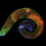
2nd Prize: Karl Norris
Spermatogenesis in a Drosophila melanogaster testis: in contact with hub cells (testis tip, violet: Armadillo), the somatic and germline stem cells give rise to gonialblasts that undergo mitosis (green: Vasa), meiosis and spermiogenesis. The latter two stages are stained in red (Fmr1), nuclei are stained by DAPI (blue).
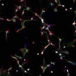
3rd Prize: Drinalda Cela
This fluorescence microscopy image shows neutrophil extracellular traps (NETs), produced in response to haem and TNF stimulation. NETs are composed of DNA (in blue), granule proteins (such as neutrophil elastase, in red) and histones (in green). When neutrophils die via NETosis they release these web-like structures, which trap microbes and stimulate additional immune responses.
You can read more about the people behind these images here.