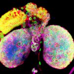
1st prize: Chantal Roubinet
Psychedelic brain: these beautiful brain lobes from Drosophila larvae illustrate the diversity of the cells that work together to generate a functional brain. This confocal image shows the nuclear envelope of nuclei in green (Lamin), the chromatin in blue (DAPI), Tubulin in Magenta and a cortical marker in red (dMoesin)
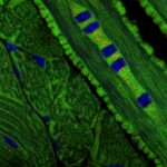
2nd prize: Alan Prescott
Mitochondrial organisation and turnover in the tongue revealed by the mitoQC mouse model Images taken from a frozen section of the tongue from the mitoQC mouse. Mitochondria are labelled with GFP(Green) and mCherry(Red). Nuclei are blue.
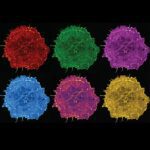
3rd prize: Hoang Anh Le Drops of colour. This is a still image from a live imaging movie of a COS-7 cell expressing the marker LifeAct showing the intricate network of the actin cytoskeleton. The image is inspired by the Pop Art style picture of Marilyn Monroe by Andy Warhol.
You can read more about the people behind these images here.