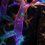
1st place: Liam Hill
This structure is the bronchial tree of an adult mouse that’s cleared by FLASH and stained for smooth muscle actin. This stain highlights the bands of smooth muscle that surround the airways modulating airflow into and out of the lung. Maximum intensity projection of 3D image. Colour corresponds to Z-depth.
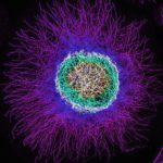
2nd place: Nikki Paul
Microtubules in a B16F1 melanoma cell imaged using a Zeiss Elyra 7 using Structured Illumination Microscopy (SIM). Cells were fixed and stained prior to imaging using a 63x oil objective. Optimal Z-sectioning was acquired followed by SIM processing, and a colour-coded projection of the Z-stack was applied using Zen software.
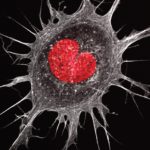
3rd place: Hoang Anh Le
At the heart of a cancer cell. The image is an Ewing’s sarcoma cell with a peculiar heart-shaped nucleus stained with DAPI (red) and the actin cytoskeleton is visualised with phalloidin (white). Image taken using the Airyscan Zeiss 880 system.
At the heart of a cancer cell. The image is an Ewing’s sarcoma cell with a peculiar heart-shaped nucleus stained with DAPI (red) and the actin cytoskeleton is visualised with phalloidin (white). Image taken using the Airyscan Zeiss 880 system.
You can read more about the people behind these images here.